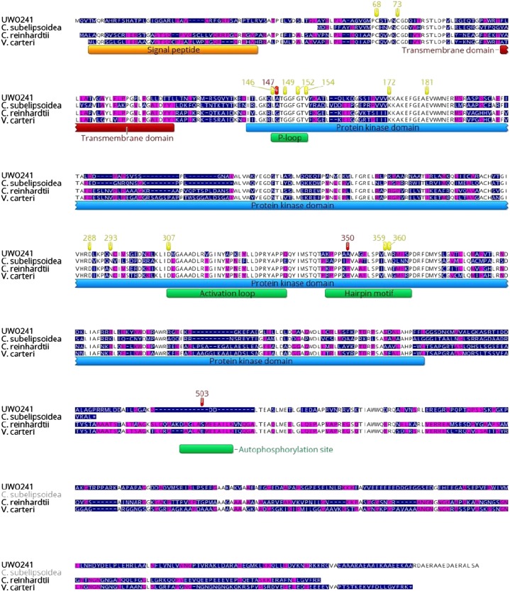
Figure 3.
The Stt7 protein of Chlamydomonas sp. UWO241 is aligned with those of C. subellipsoidea, C. reinhardtii, and V. carteri. The color of the residue depends on the level of conservation among species (white, fully conserved; yellow, 80–100% similar; purple, 60–80% similar; blue, <60% similar). Regions corresponding to the putative signal peptide, transmembrane domain, and kinase domain are indicated. The positions of important sequence patterns are marked with a green box, and highly conserved residues are highlighted in yellow (according to Guo et al. [2013] and Lemeille et al. [2010]).

