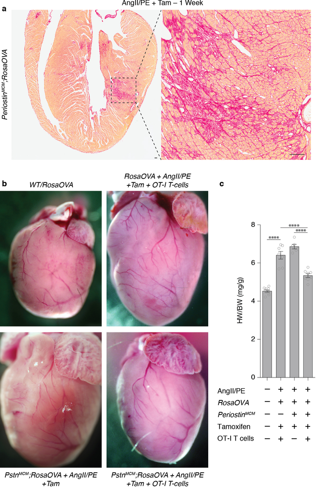
Extended Data Fig. 1.
Cardiac fibrosis and hypertrophy.
(a) Pircro-Sirius Red staining for cardiac fibrosis (red) in a heart coronal section from a PeriostinMCM;RosaOVA mouse treated with AngII/PE and tamoxifen for 1 week. High powered field of left ventricular free-wall (right). Representative image of 3 biologically independent animals with similar results. (b) Control and experimental hearts were measured (weight, mg) and images captured. (c) Quantification of heart weight to body weight (HW/BW) ratio of indicated genotypes and conditions (mean ± SEM). ****P < 0.0001 (one-way ANOVA between groups P < 0.0001; post-hoc multiple comparisons, Tukey’s test, n = 10, 7, 6, 8 biologically independent animals, respectively). Scale bars = 100μm.

