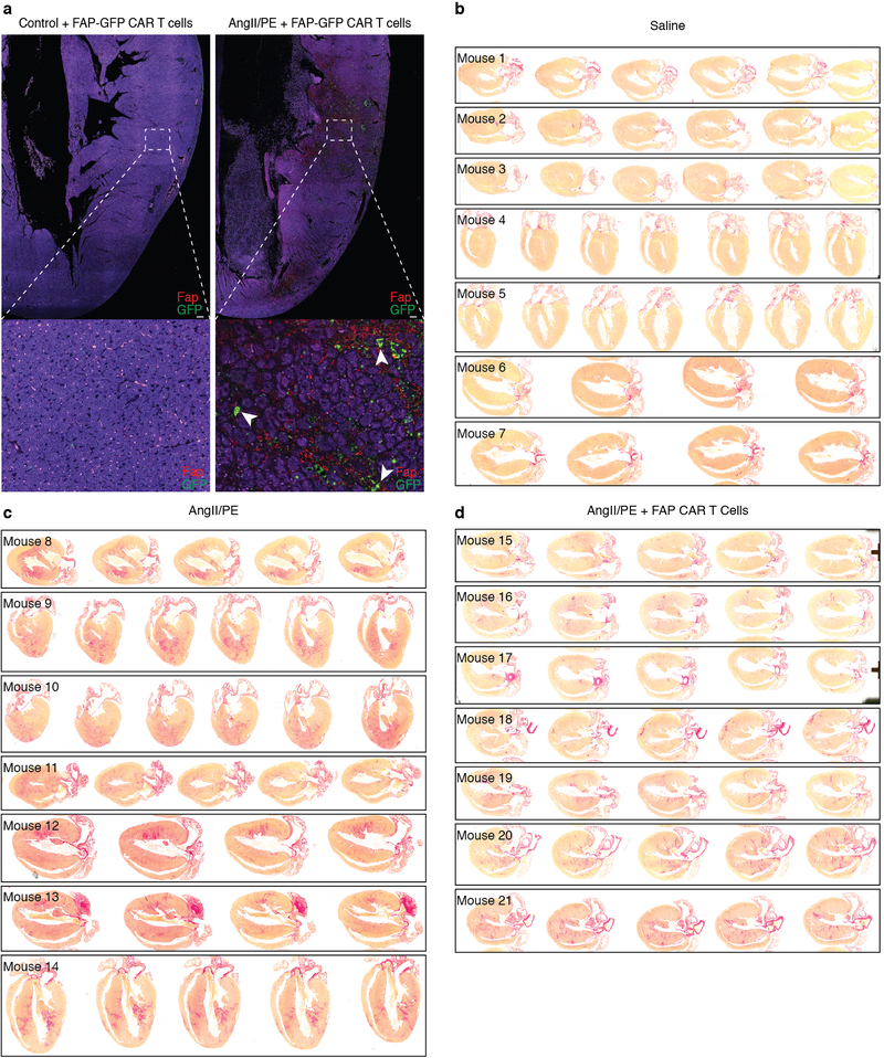
Extended Data Fig. 4.
FAP CAR T cells infiltrate the heart and reduce cardiac fibrosis.
(a) Immunohistochemistry for Fap (red) and GFP (green) on the left ventricular free wall of mouse heart coronal sections. WT C57Bl/6 mice were treated with (right) or without (left) AngII/PE for 1 week, injected with FAP-GFP CAR T cells and sacrificed 1 day later. FAP-GFP CAR T cells co-localize with FAP expressing cells (arrowheads, bottom). (b-d) Picro-Sirus Red staining of hearts from 21 individual mice (#1–21) treated for 4 weeks with either saline (b), AngII/PE (c), or AngII/PE + FAP CAR T cells (d) to assess for fibrosis (red). Representative images of 2 independent experiments with similar results. Scale bars = 100μm.

