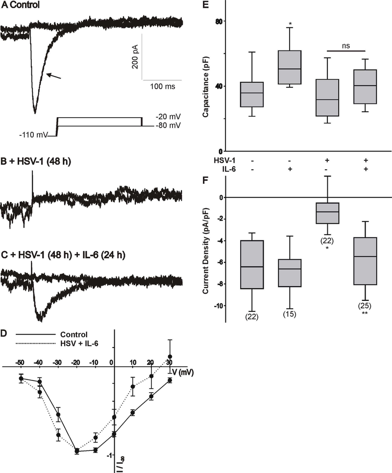
Figure 1.
Changes in whole cell Ca2+ currents in differentiated ND7/23 cells following HSV-1 infection and treatment with IL-6. A-C) Example of whole cell T-type Ca2+ currents generated in a differentiated ND7/23 cell following HSV-1 infection and treatment with IL-6. Differentiated ND7/23 cells were infected overnight with HSV-1. After a 24 h infection period, cells were treated with IL-6 (20 ng/mL) for another 24 h. Recordings from control (non-treated) and treatment groups were performed at the end of the 48 h period. In this and subsequent figures, the voltage step protocol is shown below the current trace. Note that the transient component (arrow) generated by voltage step to −20 mV from a holding potential of −110 mV was eliminated following HSV-1 infection (A,B). Stimulation with IL-6 reverses the inhibitory effect of HSV-1 infection on T-type Ca2+ currents (B,C). Scale bars in A are the same for B and C. D). Normalized current-voltage (I-V) relationship generated by the activation of T-type Ca2+ channels in differentiated ND7/23 cells (non-infected vs. HSV+IL-6 treated cells). Current amplitudes at different voltages were normalized to that generated by a voltage step to −20 mV from a holding potential of −110 mV (I/I −20). E) Comparison of cell capacitance in ND7/23 cells following HSV-1 infection and treatment with IL-6. The number of cells recorded under each condition is presented in parenthesis in panel F. F) Mean T-type Ca2+ current densities generated in ND7/23 cells following HSV-1 infection and treatment with IL-6. T-type Ca2+ current density was calculated from the peak current amplitude generated by a voltage step to –20 mV from a holding potential of −110 mV. The number of cells recorded under each condition is presented in parenthesis from at least 3 different cell cultures. * denotes p ≤ 0.05 vs. control (non-treated) cells; ** denotes p ≤ 0.05 vs. HSV-1 infected cells.

