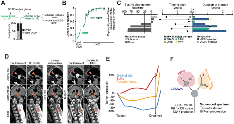
Figure 4. BRAF mutations and response to RAF/MEK/ERK-directed therapy.
A) The distribution of different BRAF mutations broken down by glioma type in the study cohort. B) The fraction of cancer cells harboring either V600E or non-V600 activating mutations in BRAF indicates GBMs harbor largely subclonal non-V600 BRAF mutations while non-GBM histologies possess predominantly clonal V600E mutations. The median and interquartile range for cancer cell fractions of BRAF V600 mutations in other BRAF-mutant cancer types are shown for context. C) On left, the best response in BRAF V600-mutated patients receiving MAPK-directed targeted therapy as measured by change in the sum of diameters across all measured lesions from initial scan. At right is the duration of therapy and date of resection and BRAF status. D) The clinical course of a single BRAF-mutant patient that progressed from low- to high-grade disease, and then experienced a durable 87% tumor regression on MAPK targeted therapy (bottommost patient in panel c). Imaging is shown for three lesions (original site of disease, posterior fossa, and spinal metastasis). RAF monotherapy was ultimately held after initial response for consolidative radiotherapy, upon which the patient clinically deteriorated and was subsequently treated with combination RAF+MEK therapy, to which they re-responded with similar lesion-specific differences in the degree of response. E) The degree of tumor regression over time is shown in the three disease sites. F) The evolutionary relationship among multiple sequenced lesions in the same patient in panels (D-E) indicated little additional genetic heterogeneity among sites except for a CDKN2A deletion specific to the non-responding spine lesion.

