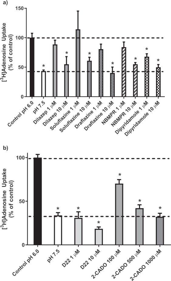
Figure 3.

Inhibition of hENT4-mediated [3H]adenosine uptake. Uptake of 1 µM [3H]adenosine by PK15-hENT4 cells (6 min incubation) was assessed at pH 6.0 in the presence and absence of the indicated concentrations of established inhibitors of ENT1 and ENT2 (Panel a), or the hENT4 inhibitor D22 and the adenosine analogue 2-chloroadenosine (2-CADO) (Panel b). Uptake in the absence of inhibitors at pH 7.5 is also shown. Data are expressed as a percentage of the uptake measured at pH 6.0 in the absence of inhibitor (Control). Each bar is the mean ± S.E.M. from 5 experiments conducted in duplicate. *Significantly different from Control (One way ANOVA with Dunnett’s multiple comparisons test, P < 0.05).
