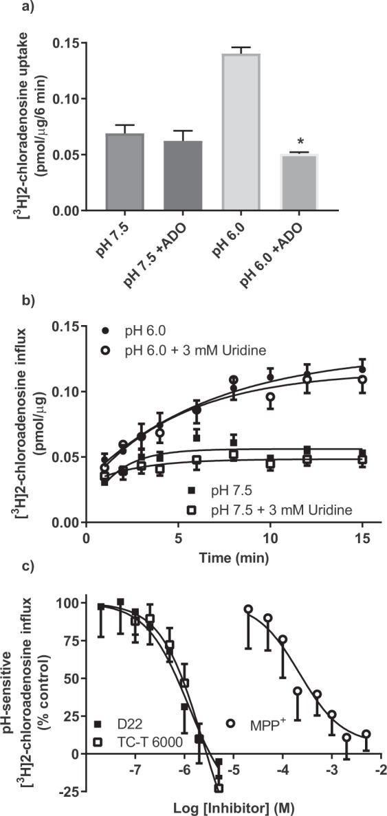
Figure 6.

Inhibition of [3H]2-chloroadenosine influx in PK15-hENT4 cells. (a) The uptake of 30 µM [3H]2-chloroadenosine (6 min incubation) was assessed at pH 7.5 and pH 6.0 in the presence and absence of 10 mM adenosine (ADO). Each bar represents the mean ± S.E.M. from 5 experiments conducted in duplicate. *Significant difference ± ADO at pH 6.0 (One way ANOVA with Tukey’s multiple comparisons post-test, P < 0.05). (b) The uptake of 30 µM [3H]2-chloroadenosine was assessed at the indicated times at pH 7.5 and pH 6.0 in the presence and absence of 3 mM uridine (ADO). Each point represents the mean ± S.E.M. from 5 experiments conducted in duplicate. Note that there is no significant effect of uridine on 2-chloroadenosine uptake at either pH 7.5 or pH 6.0. (c) The uptake of 30 µM [3H]2-chloroadenosine (6 min incubation) was assessed at pH 6.0 in the presence and absence of the indicated concentrations of D22, TC-T6000 or MPP+. Data are shown as % of control where 100% was the uptake at pH 6.0 in the absence of inhibitor and 0% was the uptake measured at pH 7.5 in the absence of inhibitor. Each point represents the mean ± S.E.M from 5 (D22, TC-T6000) or 6 (MPP+) experiments conducted in duplicate. Data shown were fitted by sigmoid curves with the maximum constrained to 100% but with no constraint on the minimum.
