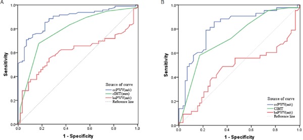
Fig. 2.

ROC curves of three methods for diagnosing CMSA and its early stage
A. ROC curves of ccPWV, cIMT, and baPWV for evaluating CMSA. The AUCs of the three methods are significantly different (P ≤ 0.001). B. ROC curves of ccPWV, cIMT, and baPWV for detecting early stage CMSA. The AUCs of the three methods are significantly different (P < 0.001). ROC, receiver operating characteristic; CMSA, the segment atherosclerosis between CCA and the ipsilateral MCA; AUC, area under the curve; ccPWV, carotid–cerebral pulse wave velocity; cIMT, carotid intima–media thickness; baPWV, brachium-ankle pulse wave velocity.
