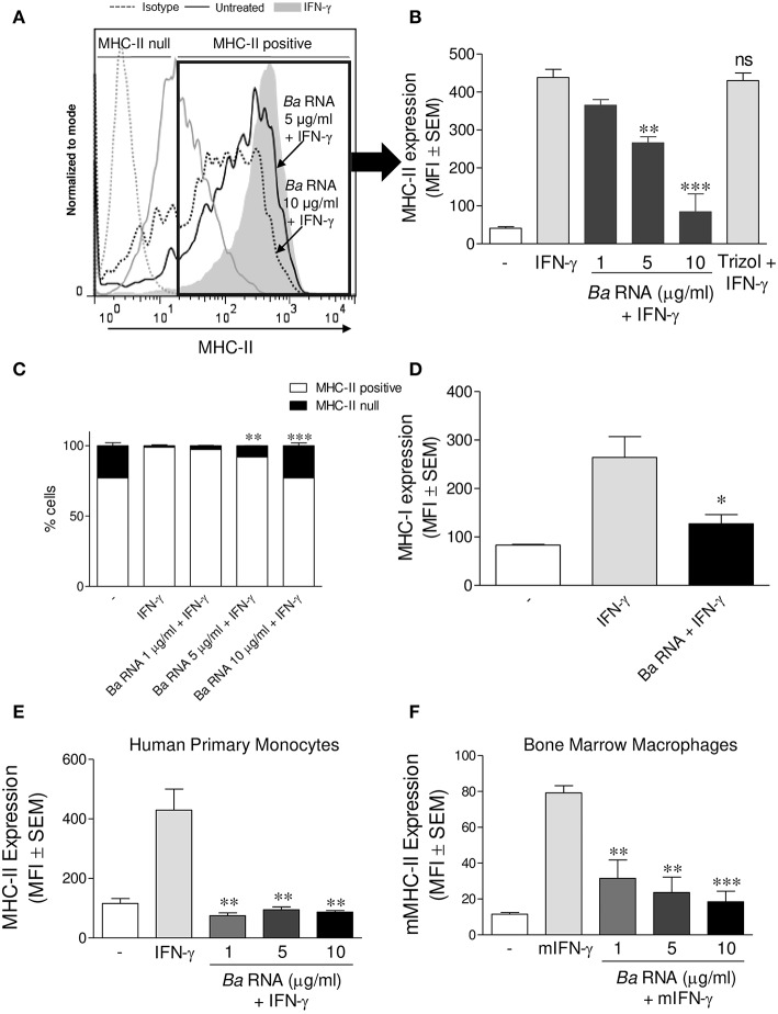
Figure 1.
B. abortus RNA down-modulates MHC-II on monocytes/macrophages. (A,B) THP-1 cells were treated with different doses of B. abortus RNA in the presence of IFN-γ for 48 h. MHC-II expression was evaluated by flow cytometry. (A) Flow cytometry histograms (showing MHC-II positive and null cells) representative of bars showed in (B). (B) Bars represent the arithmetic means ± SEM of MHC-II positive cells corresponding to five independent experiments. Trizol extracted products in the absence of bacteria were used as a control. (C) Quantification of cells expressing MHC-II (MHC-II positive cells) or not (MHC-II null). Data is expressed as the percentage of cells ± SEM of three independent experiments. (D) THP-1 cells were treated with B. abortus RNA (10 μg/ml) in the presence of IFN-γ for 48 h. MHC-I expression was evaluated by flow cytometry. (E,F) Peripheral blood-purified monocytes (E) and bone marrow macrophages (F) were stimulated with different doses of B. abortus RNA in the presence of IFN-γ for 48 h. MHC-II expression was assessed by flow cytometry. Bars represent the arithmetic means ± SEM of five independent experiments. MFI, mean fluorescence intensity; mIFN-γ, murine IFN-γ. ns, non-significant. *P < 0.05; **P < 0.01; ***P < 0.001 vs. IFN-γ-treated cells.

