Figure 9.

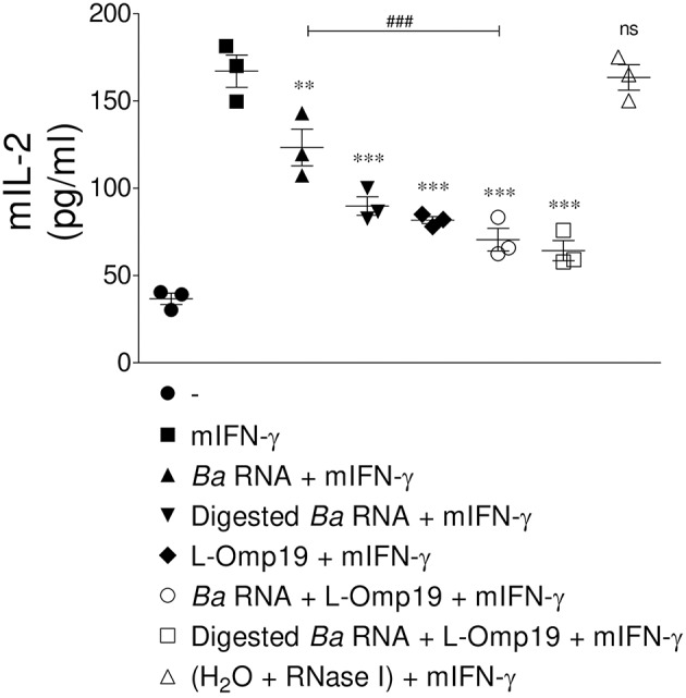
MHC-II down-modulation correlates with diminished antigen presentation to CD4+ T cells. BMM were treated with B. abortus RNA (10 μg/ml), digested B. abortus RNA, L-Omp19 (1 μg/ml) or their combination in the presence of mIFN-γ for 48 h. Then, cells were washed and incubated with 100 μg/ml of OVA peptide for 3 h at 37°C. Afterwards, cells were washed and co-cultured for 20 h at 37°C with BO97.10 cells, a T cell hybridoma specific for OVA peptide. T cell activation was measured by quantifying mIL-2 secretion in culture supernatants. **P < 0.01; ***P < 0.001 vs. mIFN-γ-treated cells; ns, non-significant; ###P < 0.001 vs. Ba RNA.
