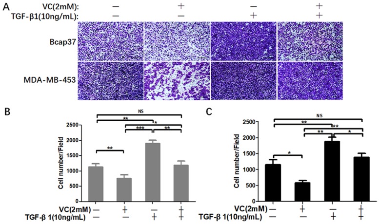
Figure 6.
High-dose VC inhibited cell invasion promoted by TGF-β1 in breast cancer cells. (A) Breast cancer cells were seeded in the upper chamber, and then cells were incubated with 2 mM VC and/or 10 ng/mL TGF-β1 for 24 h. Cancer cells invaded the underside of the membrane were fixed in 1% methanol and stained by crystal violet. Quantification of invaded Bcap37 (B) and MDA-MB-453 (C) cells were counted in transwell invasion assays. *p<0.05, **p<0.01 and ***p<0.001.

