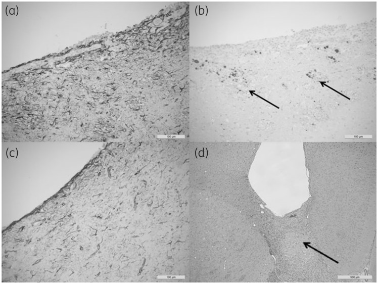
Figure 2.
(a) Section showing the extent of reactive gliosis in the tract wall (GFAP) for a control catheter after 1 week. The surrounding tissue gliotic response, demonstrated in both the control and impregnated groups after 1 week, reflected surgical trauma at this site, which subsided after 4 weeks. (b) Stains for β-APP revealed traumatic axonal injury (deposits are indicated by arrows) within the walls of the tracts related to surgical trauma. This was present in all groups and varied in extent, but was independent of the contents of the catheter. (c) The gliotic response at 4 weeks (GFAP) was reduced in both the control and impregnated catheters. The traumatic axonal injury resulting from the catheter implant persisted at 4 weeks, but the extent of inflammation at 4 weeks was proportional to the degree of traumatic injury and there was no subjective evidence that the impregnated catheter induced either inflammation or gliosis in excess of that produced by the control catheter. (d) An example of a catheter (control) at 4 weeks (β-APP) showing an inflammatory infiltrate extending from the catheter tip to traverse the corpus callosum (arrow), demonstrating significant axonal pathology in the corpus callosum, which presumably resulted in traumatic vascular injury (not shown). These cases showed tract inflammation at 4 weeks regardless of the contents of the catheter segment tip.

