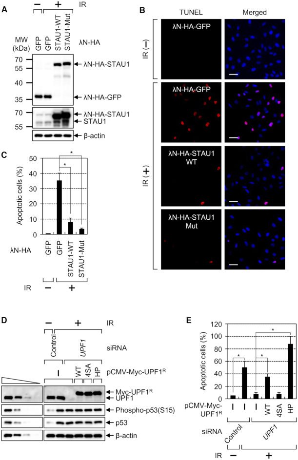
Figure 8.
IR-induced apoptosis is significantly inhibited by increased CBC replacement by eIF4E via either STAU1 overexpression or UPF1 downregulation. (A–C) The TUNEL assay for monitoring IR-induced apoptosis. HeLa cells transiently overexpressing λN-HA-GFP, λN-HA-STAU1-WT or λN-HA-STAU1-Mut were either untreated or exposed to IR and then analyzed by the TUNEL assay. (A) Western blots revealing comparable expression of λN-GFP, λN-STAU1-WT or λN-STAU1-Mut. (B) Representative images of the TUNEL assay. Scale bar, 50 μm. (C) Quantitation of apoptotic cells. The stained cells (apoptotic cells) were counted, and the ratios are presented as a percentage (%). n = 3; *, P < 0.05. (D and E) The complementation experiment with siRNA-resistant UPF1. HeLa cells either undepleted or depleted of endogenous UPF1 were transiently transfected with a plasmid expressing Myc-UPF1R-WT, -4SA or -HP. Then, the cells were either not treated or exposed to IR. (D) Western blots revealing specific downregulation and comparable expression of Myc-UPF1R-WT or mutant relative to the level of endogenous UPF1. (E) Quantitation of apoptotic cells. The stained cells (apoptotic cells) were counted, and the ratios are presented as a percentage (%). n = 2; *, P < 0.05.

