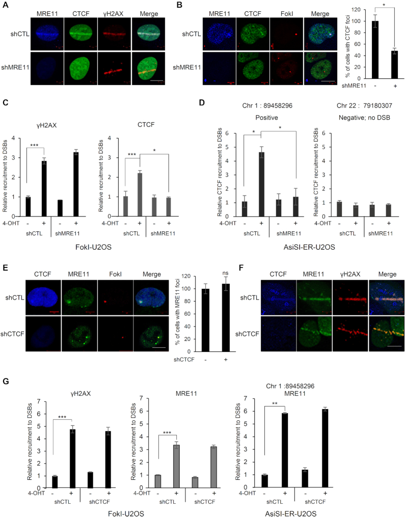
Figure 2.
MRE11-dependent recruitment of CTCF at sites of DNA damage. (A) MRE11-depleted (shMRE11) or control (shCTL) U2OS cells were presensitized with BrdU and subjected to laser micro-irradiation. Cells were fixed and stained with the indicated antibodies; scale bar: 10 μm. (B) Immunofluorescence was performed 4 h after induction of double-strand breaks by ER-mCherry-lacR-FokI-DD in the FokI-U2OS reporter cells transfected with shRNA targeting MRE11 (shMRE11) or control shRNA (shCTL); scale bar: 10 μm. The plot represents the percentage of cells positive for CTCF co-localized at mCherry-FokI foci. Data are the means ± SD of three independent experiments. More than 100 cells were counted in each experiment; *P≤ 0.05. (C) ChIP-qPCR was performed with an antibody to γ-H2AX or CTCF in the FokI-U2OS DSB reporter cells transfected with the indicated shRNA (shCTL as a control or shMRE11 targeting MRE11), with (+) or without (−) induction of DSBs by mCherry-LacI-FokI. The values of recruitment to DSBs were relative to those of cells without the induction of DSBs. All qPCR reactions were performed in triplicate, with the SEM values calculated from at least three independent experiments; *P≤ 0.05; ***P≤ 0.001. (D) AsiSI-ER-U2OS cells were transfected with the indicated shRNA, with (+) or without (−) induction of DSBs by AsiSI. CTCF and chromatin were immunoprecipitated with anti-CTCF antibody. The fold enrichment values were relative to those of cells without induction of DSBs. Primers on chromosome 22 (no DSB) were used as negative controls. Data are the means ± SD of at least three independent experiments, and all qPCR reactions were performed in triplicate. (E) Immunofluorescence was performed 4 h after induction of double-strand breaks (DSBs) by mCherry-LacI-FokI in the CTCF-depleted (shCTCF) or control (shCTL) FokI-U2OS cells; scale bar: 10 μm. Bar graph represents the percentage of cells positive for MRE11 co-localized at mCherry-LacI-FokI foci. Data are the means ± SD of three independent experiments. More than 100 cells were counted in each experiment. ns, not significant. (F) Recruitment of MRE11 (green) to DSBs induced by laser micro-irradiation in the CTCF-depleted (shCTCF) and control (shCTL) U2OS cells; scale bar: 10 μm. (G) ChIP-qPCR was performed with an antibody to γ-H2AX or MRE11 in FokI-U2OS cells (left and center), and AsiSI-ER-U2OS cells (right) transfected with the control (shCTL) or CTCF shRNA (shCTCF), with (+) or without (−) induction of DSBs by FokI (FokI-U2OS) or AsiSI (AsiSI-ER-U2OS). The fold enrichment values were relative to those of cells without induction of DSBs. Data are presented as means ± SD of three independent experiments, and all qPCR reactions were performed in triplicate; **P≤ 0.01; ***P≤ 0.001.

