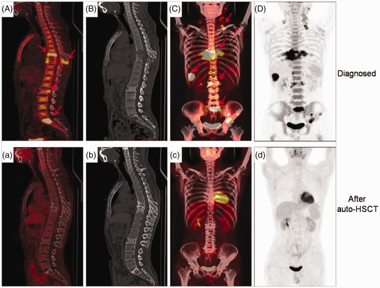
Figure 2.
The PET-CT images of a 28-year-old male patient with LCH before (1 April 2016) and after (1 January 2017) receiving auto-HSCT. (A–D) Images showing multiple lesions (arrowheads in A and C) in liver, lymph nodes, and bones, as well as osteolytic lesions (T7, T8, T9, and S1) with SUVmax 10.5 before therapy. (a–d) Images showing no highly FDG-avid lesions in liver, lymph nodes, or bones and smaller osteolytic lesions. Compared with the initial diagnosis, we found evidence of peripheral sclerosis (T7, T8, T9, and S1), an indicator of bone regeneration. PET-CT, positron emission tomography–computed tomography; LCH, Langerhans cell histiocytosis; auto-HSCT, autologous hematopoietic stem cell transplantation; SUVmax, maximum standardized uptake value; FDG, 18F-fluorodeoxyglucose.

