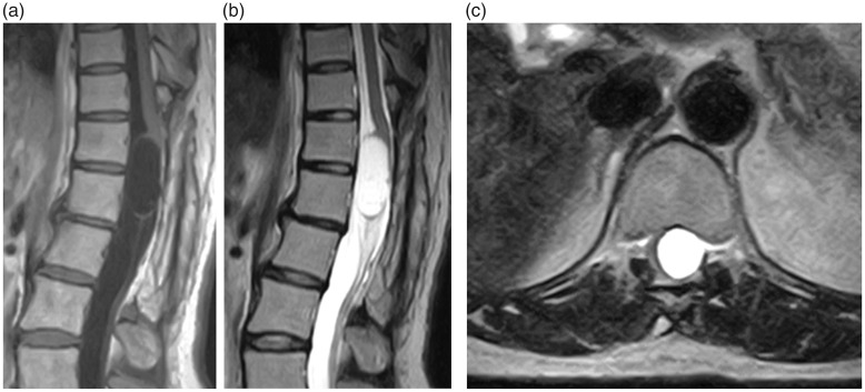
Figure 1.
(a) A T1-weighted sagittal thoracic magnetic resonance imaging section showing an intradural, extramedullary low-signal-intensity mass involving the T12–L1 level. (b) A T2-weighted sagittal section showing a mass exhibiting uniform high signal intensity. (c) A T2-weighted transverse section showing that the spinal cord had been pushed to the right by a high-signal-intensity mass at the T12 level.

