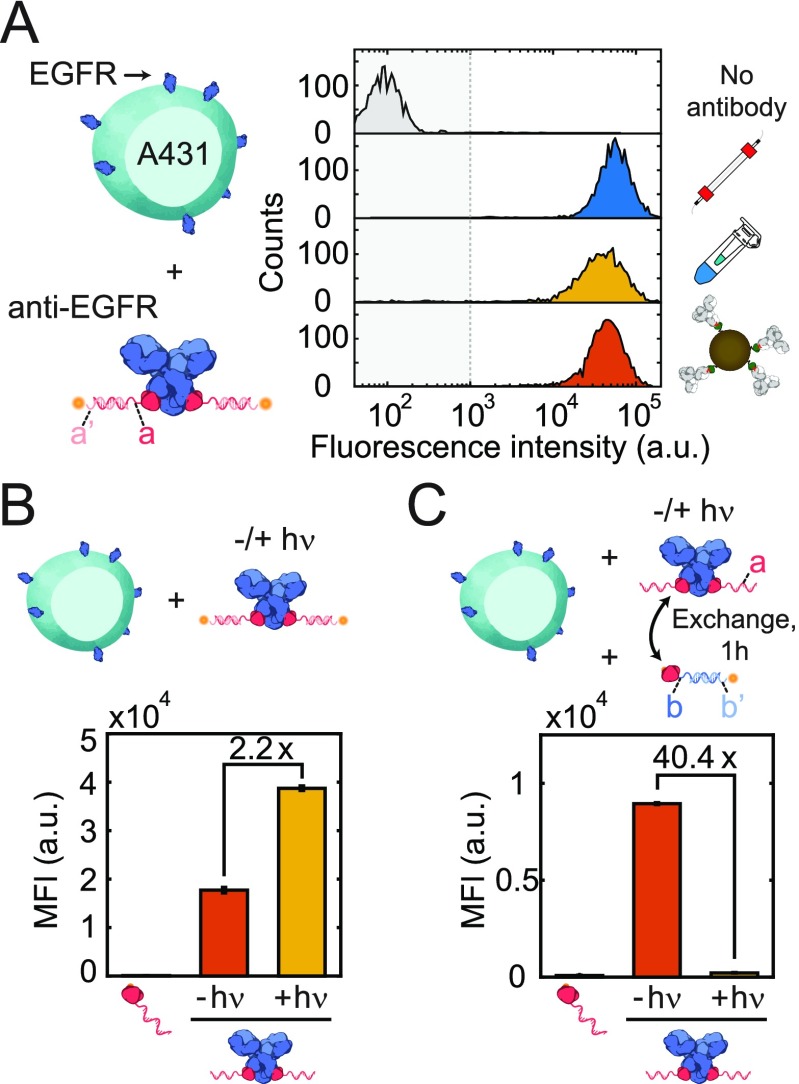
Figure 3.
Flow cytometric analysis of EGFR-expressing A431 cells using 10 nM pG-ODN-functionalized Cetuximab, a, hybridized to a CY5-functionalized ODN, a′. (A) Antibody activity after purification using SEC, ultrafiltration, and magnetic beads. The fluorescence intensity of pG-ODN-Cetuximab-labeled A431 cells was compared with that of A431 cells incubated with only pG-ODN. (B) Labeling efficiency and (C) cross-contamination of pG-ODN noncovalently (−hν) or covalently (+hν) coupled to Cetuximab. pG-ODN-functionalized Cetuximab was incubated for 1 h with a 20 mol equiv of a competing pG-ODN sequence. MFI represents the median fluorescence intensity, and error bars represent the SD (n = 3).

