
Figure 5.
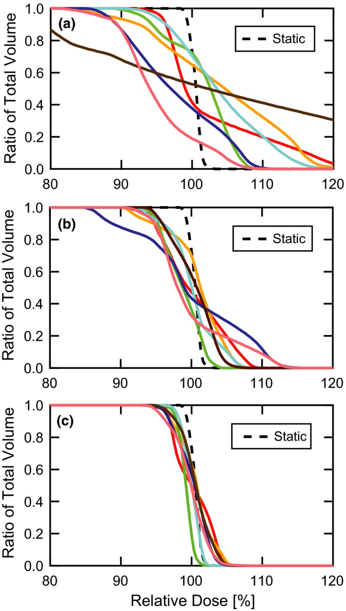
DVH comparisons. DVH curves are shown for (a) target motion without motion compensation, (b) target motion with a gate threshold of 0.3 cm, and (c) target motion with a gate threshold of 0.3 cm and maximum‐MU repainting with 0.004 MU per spot. The plots contain seven distinct patient breathing patterns along with the static target for reference. The target moves in the plane of the beam’s eye view, orthogonal to the primary scanning axis of the proton delivery system. DVH, dose volume histograms.