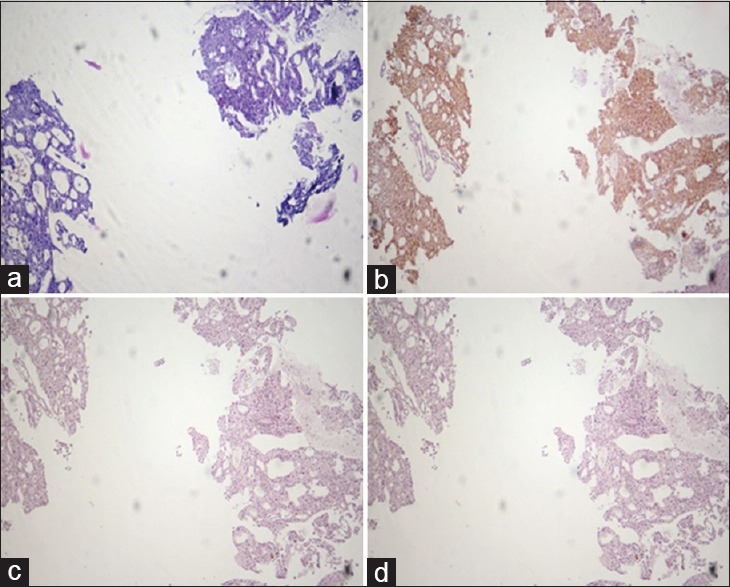
Figure 1.

Carcinoma prostate: poorly differentiated. Comparison of hematoxylin and Eosin (a), AMACR (b), HMWCK (c), and P63 (d). Staining in serial sections of a small focus of prostatic Adenocarcinoma. (b) Diffuse intense cytoplasmic staining of the neoplastic glands with AMACR. The diagnosis of prostatic adenocarcinoma was confirmed by the negative basal cell staining with both HMWCK (c) and P63 (d). (×10)
