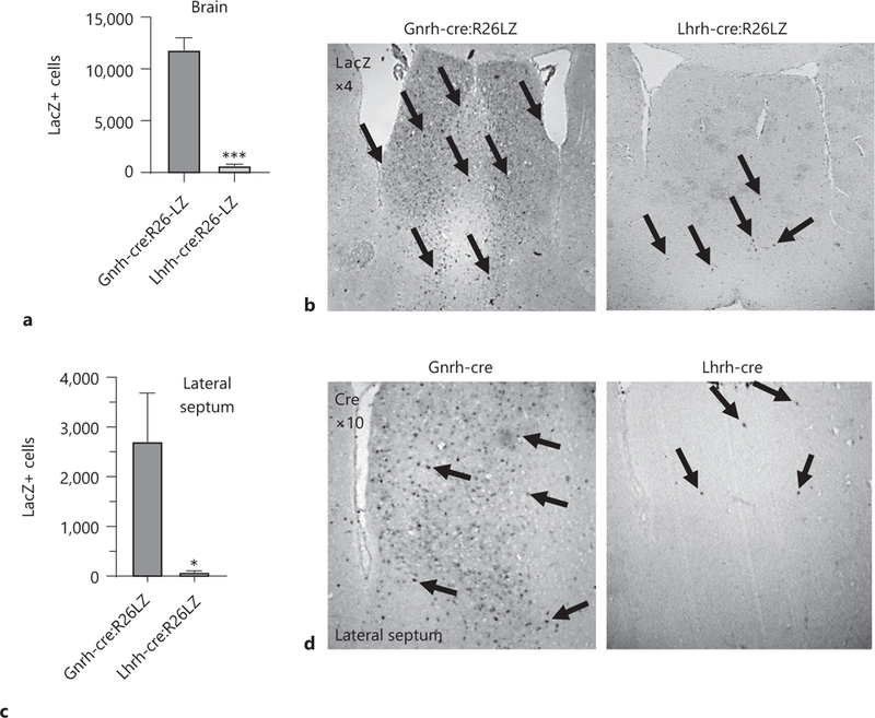
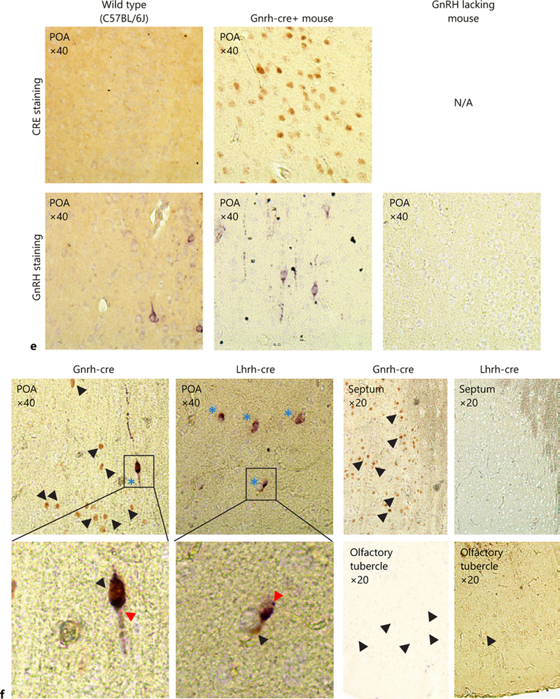
Fig. 1.


Quantification of LacZ positive (LacZ+) cells in the whole brain (a) and the lateral septum (c) from adult Gnrh-cre:Rosa26-LacZ and Lhrh-cre:Rosa26-LacZ mice, n = 3, Student t test; * p < 0.05. Immunohistochemical staining in the lateral septum for (b) LacZ in Gnrh-cre:Rosa26-LacZ and Lhrh-cre:Rosa26-LacZ (×4), and (d) CRE in Gnrh-cre and Lhrh-cre mice (×10). Arrows high-light LacZ or CRE expressing cells in coronal sections. e Negative controls for double immunohistochemistry, showing specificity of the anti-CRE and anti-LHRH antibodies. To assure that no cross-reaction of the antibodies occurred, the antibody validation was done following the entire double immunohistochemistry protocol where the anti-LHRH antibody was omitted for the CRE staining, and the anti-CRE antibody omitted for the GnRH staining. The GnRH-lacking mouse is the Gnrh-cre:Vax1-flox/flox mouse that we previously published as lacking GnRH expressing neurons in the hypothalamus as a negative control for the anti-LHRH antibody [11]. f Double immunohistochemistry for GnRH (purple, GnRH staining is indicated by a red arrow head) and CRE (brown, single staining is indicated by a black arrow head). Dual staining is indicated by blue stars. n = 3 adult brains.
