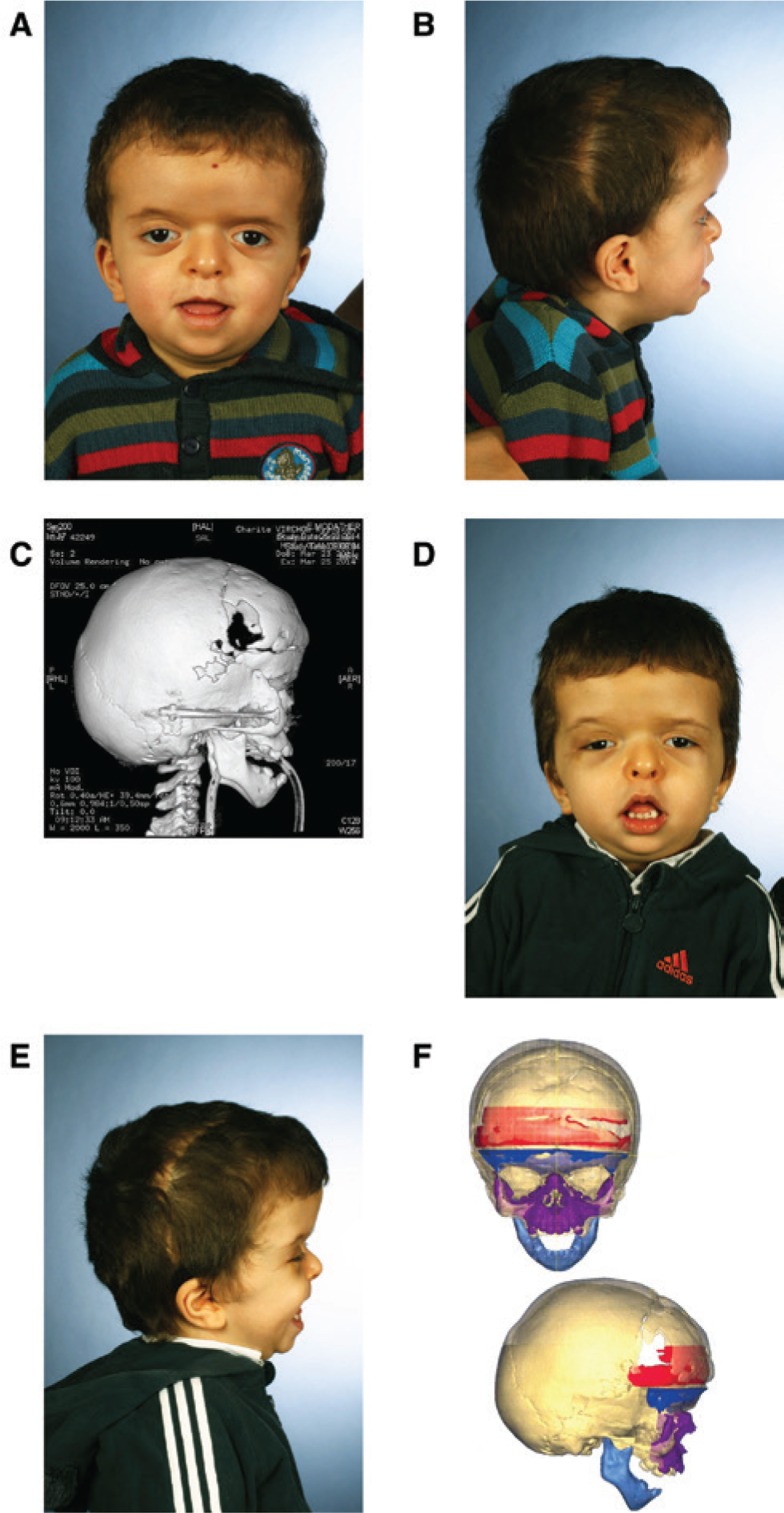
Figure 2:
3-year-old boy affected by craniofacial dysostosis (M. Apert) with midfacial retrusion.
Frontoorbital advancement with overcorrection had been performed in the first year of life (A and B). After computer-assisted frontofacial advancement of 17 mm by internal distraction devices (C), midfacial retrusion is improved (D and E). However, superimposition of predistraction and postdistraction data sets (DePuy Synthes ProPlan CMF; Materialise, Leuven, Belgium) reveals a lack of advancement in the central midface (F; violet zone), which is known to be one of the drawbacks of internal craniofacial distraction devices requiring additional corrective surgery.

