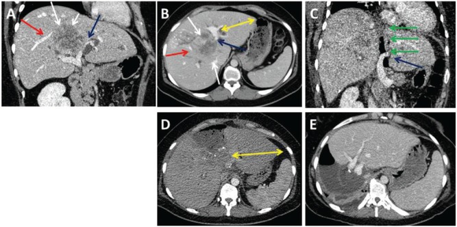
Figure 2:
A 32-year-old female patient with intrahepatic CCC.
The tumor (A and B, white arrows) had contact with a major branch of the right hepatic (A and B, red arrows) and left portal vein (A and B, blue arrows) and caused bilateral cholestasis. The ALPPS procedure was performed for curative resection. CT on POD1 after the first step demonstrates the patent left portal vein (C, blue arrow) and the dissection line along the falciforme ligament (C, green arrows). Due to the infiltration of the hilar bifurcation, the left bile duct was resected and a hepaticojejunostomy was performed for reconstruction. CT control on POD7 after the first step reveals sufficient hypertrophy of the left lateral sector (D, yellow arrows). The completion of ALPPS was done on POD7. CT 7 days after ALPPS completion displays hypertrophy of the left lateral lobe (E). Final histology confirmed a T3, N0 (0/3), G2, R0 intrahepatic CCC (∅7.5 cm).

