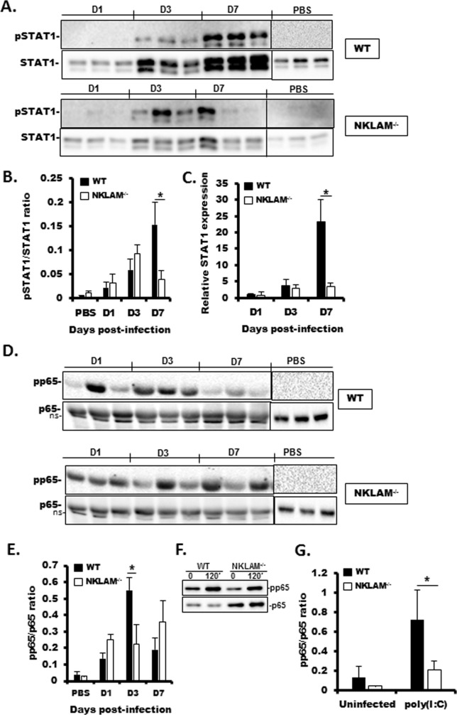
Fig 4. SeV-induced STAT1 protein expression and STAT1 and NFκB p65 phosphorylation are lower in NKLAM-/- than in WT mice.
WT and NKLAM-/- mice were infected with 500 pfu SeV/gram body weight. A) Equal amounts (20 μg) of lung homogenate protein were then probed for pSTAT1(701) and STAT1. B) Graphical depiction of pSTAT1/STAT1 densitometric ratio from A. Quantitative PCR was used to determine the relative expression of STAT1 in infected lungs (C). The mRNA levels (mean ±SD) are expressed relative to PBS-treated mice. * p < 0.05; n = 3 mice per group. D) Equal amounts of lung homogenate protein were probed for phospho-NFκB p65 (S536) and p65. E) Graphical depiction pp65/p65 densitometric ratio of D. Each lane of Western blot data represents a single mouse. * p < 0.05; comparing NKLAM-/- and WT mice at each day post-infection. n = 3 mice per group. ns; non-specific band. F) Isolated lung fibroblasts were treated with 100 μg/ml poly(I:C) for 120 min. Whole cell lysates were immunoblotted for pp65 and p65. G) Graphical representation of pp65/p65 densitometric ratio from F. Data are representative of 3 independent experiments. * p ≤ 0.05.

