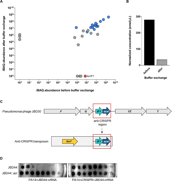
Figure 7. Anti-CRISPR AcrIF1 is not incorporated into the JBD30 virion.
(A) Analysis of absolute protein abundance of phage proteins before and after buffer exchange. Phage particle proteins are highlighted in blue. AcrIFI is shown in red. (B) Estimated concentration of AcrIF1 in phage particles before and after additional purification by buffer exchange. (C) Schematic representation of the genomic context of AcrIF1 from phage JBD30. The anti-CRISPR region (outlined red) was inserted into a transposon, which was used to randomly introduce the anti-CRISPR region into phage JBD44. F and G encode phage head and tail morphogenesis proteins, respectively. I/Z encodes the protease/scaffold and T encodes the major head protein. (D) Tenfold dilutions of lysates of phage JBD44 and phage JBD44 carrying the JBD30 anti-CRISPR locus (JDB44::acr) were applied on lawns of PA14 and PA14 CRISPR expressing a crRNA targeting phage JBD44 (JBD44 crRNA) from a plasmid. A representative image of at least three biological replicates is shown. See also Table S1.

