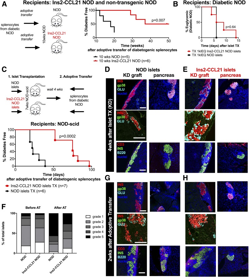
Figure 5.
Local CCL21 expression by β-cells is sufficient to induce FRC-like cell–containing TLOs and mediate systemic protection from diabetes. A: ATs of splenocytes from diabetic nontransgenic NOD mice into 10-week-old Ins2-CCL21 NOD mice (red, n = 6, median survival time 48 weeks, P = 0.007) or into age-matched nontransgenic mice (black, n = 5, median survival time 18 weeks). B: Percent of diabetes-free spontaneously diabetic NOD mice transplanted (epididymal fat pad) with 1,000 islet equivalents (IEQ) islets from Ins2-CCL21 NOD (red, n = 4) or nontransgenic (black, n = 1) NOD mice. Experimental details are reported in Table 2. C: AT of splenocytes from recently diabetic NOD mice into 7- to 11-week-old NOD-scid mice transplanted (TX) in the kidney capsule (KD) with a marginal dose (500 IEQ/mouse) of islets from either Ins2-CCL21 NOD (red, n = 7, median survival time 22.5 days, P = 0.0002) or nontransgenic (black, n = 6, median survival time 75 days) mice 4 weeks prior to injection. Confocal microscope images of KD islet graft sections (KD graft) (left) and of pancreatic sections (right) of 7-week-old NOD-scid mice that received either nontransgenic (D) or Ins2-CCL21 transgenic (E) NOD islets 4 weeks before. F: Insulitis quantification (grades 1–5) of pancreatic sections of NOD-scid mice before (left) and after (right) AT of diabetic splenocytes. Mice had received a transplant of either NOD (n = 3 for both timepoints) or Ins2-CCL21 NOD (n = 3 after transplant and n = 4 after AT) islets under the KD 4 weeks before the AT. G and H: Confocal microscope images of KD islet graft sections (KD graft) (left) and pancreatic sections (right) of NOD-scid mice that received either nontransgenic (G) or Ins2-CCL21 transgenic (H) NOD islets followed by AT of splenocytes from recently diabetic NOD mice. Sections were stained for insulin (red), stromal cells (gp38, green), and glucagon (cyan) (top panels) or for T cells (CD3, red), insulin (green), and B cells (B220, cyan) (bottom panels). Nuclei were counterstained with DAPI (blue). Scale bar, 100 µm. wks, weeks.

