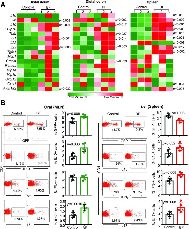
Figure 3.
Orally and systemically administered BF induces distinct immune responses. Eight-week-old female NOD mice were given HK BF for 3 consecutive days by oral gavage (500 μg/mouse/day) or by i.v. injection (10 μg/mouse/injection). A: On day 4, one set of mice was euthanized, and cDNA prepared from distal ileum and distal colon of control and BF-fed (oral) mice and spleen cells from control and BF-injected (systemic) mice were subjected to qPCR assay and the expression levels of cytokines and noncytokine factors compared. Expression levels relative to β-actin expression were plotted as heat maps using the Morpheus application. The assay was performed in triplicate (n = 3 mice/group), and for each mouse, the average values were used for each lane. This experiment was repeated using three mice/group at least once with similar statistical trends in outcomes. B: A set of NOD-Foxp3-GFP mice from a similar experiment (treated with BF for 3 days) was euthanized on day 7. MLN cells from BF-fed mice and spleen cells from BF-injected and control mice were examined for GFP+CD4+ cells or ex vivo stimulated with PMA and ionomycin for 4 h and examined for intracellular cytokines by FACS. Representative FACS plots (left) and mean ± SD of percentage of cells that are positive for specific markers (right). The assay was performed in duplicate for each mouse (n = 5 mice/group). This experiment was repeated at least twice (using three or four mice/group) with similar statistical trends in outcomes. The statistical significance was assessed by Mann-Whitney test for all panels. Supplementary Fig. 2 shows Foxp3+, IFN-γ+, IL-10+, and IL-17+ CD4 T-cell frequencies from spleen of orally treated mice and MLN of i.v.-treated mice used for panel B. Supplementary Fig. 3 shows Foxp3+, IFN-γ+, and IL-10+ CD4+ T-cell frequencies of PP, SiLP, LiLP, and spleen of mice that received BF orally and spleen and PP of mice that received i.v. injection of BF for an extended period.

