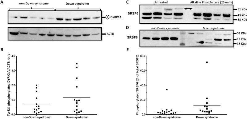
Fig. 2. Analysis of DYRK1A and SRSF6 phosphorylation status in myocardium from donors with and without Down syndrome.
(A) Representative immunoblots for Tyr321-phosphorylated DYRK1A and ACTB. (B) Tyr321-phosphorylated DYRK1A expression ratios in myocardial samples. (C) Detection of phosphorylated SRSF6 by alkaline phosphatase treatment and immunoblotting. Arrows indicate phosphorylated SRSF6 (D) Analysis of myocardial SRSF6 expression by immunoblotting. (E) Expression of phosphorylated SRSF6 in myocardial samples. Each symbol depicts the average of individual samples. Horizontal lines indicate group means. Samples were analyzed in triplicates.

