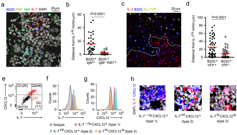
Figure 1. Location of proliferating and differentiating pre-B cells in the BM.
a, Confocal microscopy of WT BM section (8μm thick femur) stained with antibodies to IL-7 (red), B220 (blue), Ki67 (yellow), IgM (green) and DAPI (gray) to visualize the location of proliferating and IgM+ B cell progenitors. (Single color panels are presented in Supplementary Fig. 1a. The image is representative of 4 independent images from 3 WT mice) b, Distance of IgM+ and IgM−Ki67+ B cell progenitors from IL-7hif niches (white dashed line). Data were pooled from three independent experiments. Distance of each cell counted were shown with mean values (horizontal bars). P values were calculated by unpaired t-test. c, Visualization of B cell progenitors in BM sections (8μm thick femur) of Ck-YFP mice with antibodies to IL-7 (red) and CXCL12 (blue) by confocal microscopy. The image is representative of 3 independent images from 3 WT mice. d, Distances of YFP+ B cell progenitors from IL-7hi niches (white dashed line) in BM of Ck-YFP mice. Data were pooled from two independent experiments. Distance of each cell counted were shown with mean values (horizontal bars). P values were calculated by unpaired t-test. e, Flow cytometric analysis of IL-7 and CXCL12 expression by cultured BM stromal cells (n=3). f, g. Identification of stromal cells expressing IL-7 (i) and CXCL12 (j) by flow cytometry (n=3). k, Different areas of BM showing varying degrees of IL-7 and CXCL12 expressing stroma by Type 1, Type 2 and Type 3 stromal cells. Images are representative of 5 independent areas from 2 WT BM.

