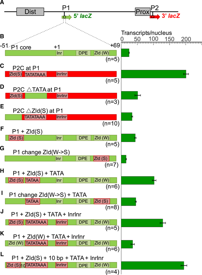
Figure 4. Testing Specific Promoter Motifs.
(A–L) Analysis of a wild type dual reporter gene (A and B) and identical reporter genes containing replacements of P1 core sequences (B) with P2 sequences (C–E), and conversions of P1 core sequences to insert specific promoter motifs from P2 (F–L). Reporter gene schematics are shown on the left and individually labeled. Complete sequences of each construct are shown in Table S3. Green bars on the right represent levels of 5′ lacZ transcripts (number per nucleus) driven by each construct. All of these constructs drive similar levels of 3′ lacZ transcripts, and we have omitted them from this figure for clarity. Error bars represent SEM. The number of embryos measured for each construct is shown below each reporter schematic.

