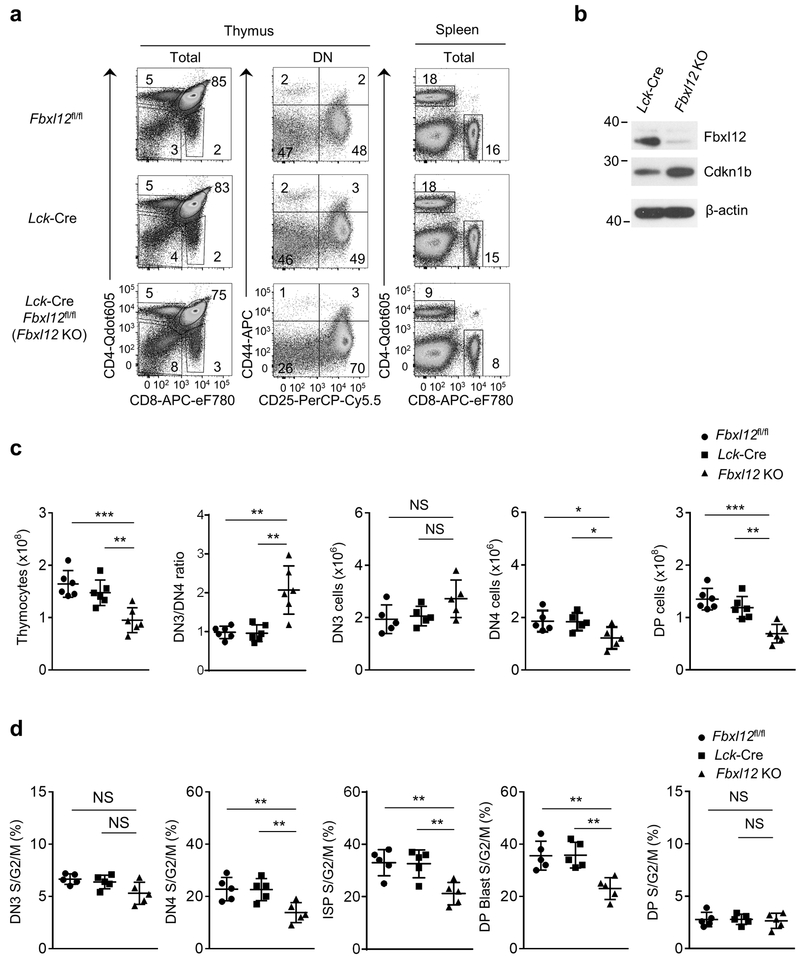
Figure 2.
Impaired β-selection-associated proliferation in Lck-Cre Fbxl12fl/fl mice. (a) Representative flow cytometry analysis showing the phenotype of thymocytes (left) or splenocytes (right) from mice of the indicated genotype. Thymus: left, CD4 vs CD8 staining of total thymocytes; center, CD44 vs CD25 staining of lineage-negative DN thymocytes. Spleen: CD4 vs CD8 staining of total splenocytes. (b) Immunoblot analysis showing absence of Fbxl12 and increased Cdkn1b expression in CD4−CD8− (DN) thymocytes from Lck-Cre Fbxl12fl/fl mice. (c) Cell numbers of the indicated thymocyte subsets and DN3/4 thymocyte ratio (n=6 mice per genotype). (d) Percentage of cycling S/G2/M stage cells in the indicated thymocyte subsets determined by staining for DAPI vs Ki-67 (n=5 mice per genotype). For all graphs, horizontal lines indicate the mean and vertical lines indicate standard deviation (±s.d.), P values were determined by unpaired two-tailed Student’s t-test. NS, not significant (P>0.05), * P<0.05, ** P<0.01, ***P<0.005. Data shown in (a) and (b) are representative of four or two independent experiments, respectively.

