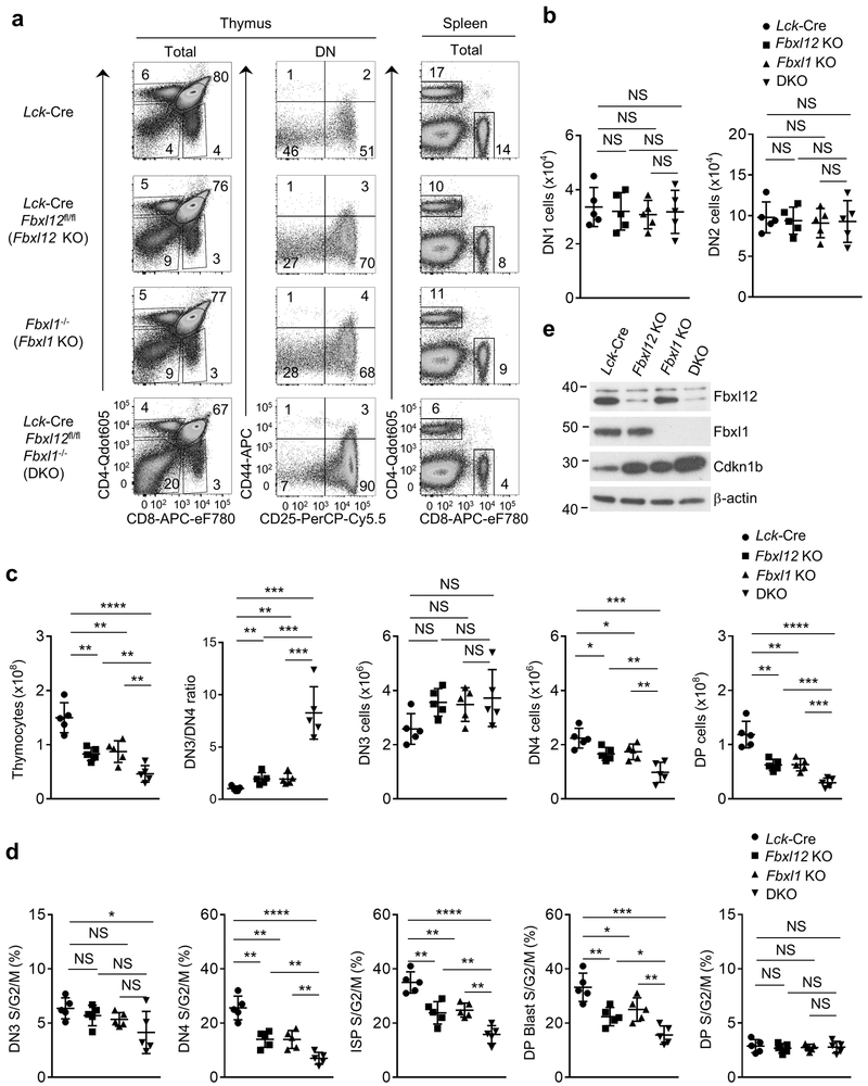
Figure 4.
Thymocyte development and β-selection-associated proliferation are strongly impaired in thymocytes lacking Fbxl1 and Fbxl12. (a) Flow cytometry analysis of cells from Thymus (left) or Spleen (right) from mice of the indicated genotype. Thymus: left, CD4 vs CD8 staining of total thymocytes; center, CD44 vs CD25 staining of lineage-negative DN thymocytes. Spleen: CD4 vs CD8 staining of total splenocytes. (b) Cell numbers of DN1 and DN2 thymocytes from mice of the indicated genoptype (n=5 mice per genotype). (c) Cell numbers of total thymocytes or the indicated thymocyte subsets and DN3/4 ratio (n=5 mice per genotype). (d) Percentage of cycling (S/G2/M) stage cells in the indicated thymocyte subsets determined by staining for DAPI vs Ki-67 (n=5 mice per genotype). (e) Immunoblot analysis showing Fbxl1, Fbxl12 and Cdkn1b expression in purified DN thymocytes from mice of indicated genotype. For all graphs, horizontal lines indicate the mean and vertical lines indicate standard deviation (±s.d.), P values were determined by unpaired two-tailed Student’s t-test. NS, not significant (P>0.05), *P<0.05, **P<0.01, ***P<0.005, ****P<0.0001. Data shown in (a) and (e) are representative of four or two independent experiments, respectively.

