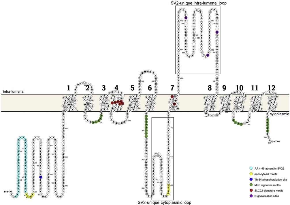
Figure 1. The SV2 proteins.
Shown is a schematic drawing of human SV2A (NCBI accession: NP_001315603) depicting the position of transmembrane domains and domains of interest. Each circle denotes an amino acid, and the orange bar the vesicle membrane. Amino terminal residues unique to SV2A and SV2C are colored green. The sequences conserved in Major Facilitator Transporters are colored magenta. Residues conserved in SV2A and SV2C (and which differ in SV2B) are colored red. Residues specific to cytoplasmic domains of SV2s are colored cyan. Tyrosine based endocytosis motifs are colored yellow. Phosphorylation and glycosylation sites are represented as dark squares. The illustration was generated using Protter tool [135]

