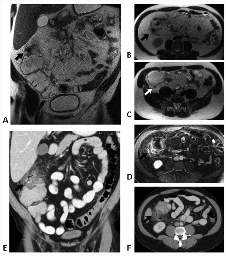
Figure 2.
52-year-old man with acute right-sided abdominal pain.
Coronal (A) and transverse (B & C) T2-weighted SSFSE images show a hypointense focus of gas and surrounding inflammatory changes involving the ascending colon (black arrows), compatible with acute diverticulitis. Note normal appendix (white arrow). Pericolonic inflammation is even more apparent on the fat-suppressed post-contrast T1-weighted images (D). Similar findings are present on the contrast-enhanced coronal (E) and transverse (F) CT images (arrows).

