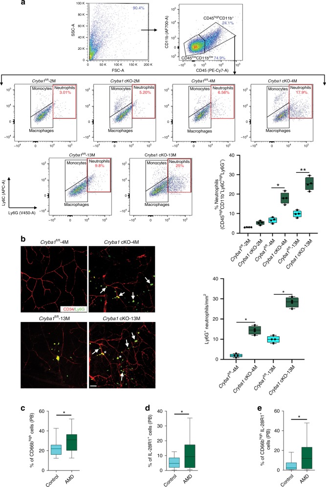
Fig. 1.
Neutrophils accumulate into the retina of Cryba1 cKO mice and in peripheral blood of human early AMD patients. a Representative dot plots are gated on the CD45+CD11b+ cells from mouse retina. The total population of CD45+CD11b+ cells is considered to be 100%, with CD45highCD11b+ (neutrophils, monocytes, and macrophages) and CD45lowCD11b+ (predominantly resident microglia) gated separately (arrows denoting population lineages). The level of Ly6C and Ly6G on the CD45highCD11b+ population was assessed to evaluate percentage neutrophils (%CD45highCD11b+Ly6ChighLy6G+ cells), which showed increased neutrophils only in 4- and 13-month-old Cryba1 cKO mouse retinas compared to aged-matched controls (Cryba1fl/fl). No differences were observed between Cryba1fl/fl and cKO retinas at 2 months of age. n = 4. *P < 0.05 and **P < 0.01 (one-way ANOVA and Tukey’s post hoc test). b Immunofluorescence studies and quantification of Ly6G+ cells (Green, Neutrophil marker) on retinal flatmounts, counterstained with CD34 (Red, marker for endothelial cells of blood vessels) revealed that neutrophils accumulated progressively in Cryba1 cKO mouse retina (white arrows) and along the retinal blood vessels (yellow, asterisk), relative to age-matched control (Cryba1fl/fl). n = 4. *P < 0.05 (one-way ANOVA and Tukey’s post hoc test). Scale bar, 50 μm. In early AMD patients, flow cytometry data revealed significant increase in the peripheral blood (PB) levels of c total neutrophils (CD66b+ cells), d total IL28R1+ cells and e IL28R1+ expressing activated neutrophils (CD66bhighIL28R1+). PB (AMD; n = 43 and Controls; n = 18). *P < 0.05 (Mann–Whitney test)

