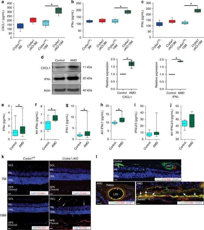
Fig. 2.
Increased levels of neutrophil regulating factors in retinas from Cryba1 cKO mice and human early AMD donor eyes. The levels of a CXCL1, b IFNα, and c IFNλ were increased in the RPE-choroid tissue homogenate of 15-month-old Cryba1 cKO mice compared to age-matched Cryba1fl/fl controls as measured by ELISA. No changes were observed in 3-month-old mice. n = 6. *P < 0.05 (one-way ANOVA and Tukey’s post hoc test). d Representative immunoblot and densitometry showed elevated CXCL1 and IFNλ in RPE lysates from early AMD donor samples compared to age-matched controls. n = 6. *P < 0.05 (one-way ANOVA and Tukey’s post hoc test). e–j Multiplex ELISA revealed significant increases in the levels of IFNα and IFNλ1 in plasma or aqueous humor (AH) of early AMD patients relative to controls. No noticeable change was observed in the plasma and AH levels of IFNλ2/3. Plasma (AMD; n = 43 and Controls; n = 18), AH (AMD; n = 6 and Controls; n = 7). *P < 0.05 (Mann–Whitney test). k Immunofluorescence assay showing increased staining of ICAM-1 (Red, neutrophil adhesion molecule) in the neural retina (white arrows) and RPE/choroid (asterisk) of aged (18-month-old) Cryba1 cKO mice compared to age-matched control. No noticeable increase in staining was observed in the retina of 7-month-old Cryba1 cKO mice. n = 5. Scale bar, 50 μm. l Immunostaining of human early AMD sections revealed increased staining of VCAM-1 (Red, neutrophil adhesion marker) in the large retinal vessels (LRV, asterisk), which were stained with CD34 (Green, marker for endothelial cells of blood vessels). Intense staining was also observed in the RPE/choroid (Yellow, white arrows). No noticeable staining for VCAM-1 was observed in the control sections. n = 5. Scale bar, 50 μm

