Fig. 7.
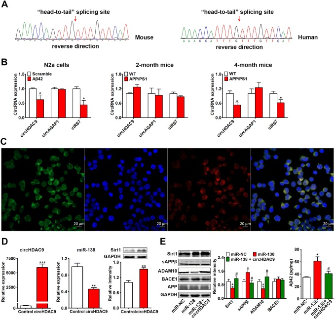
CircHDAC9 acted as a miR-138 sponge. A Sequencing of the “head-to-tail” splicing sites in mouse and human circHDAC9. B Expression of circHDAC9, circAGAP1, and ciRS-7 in N2a cells treated with Aβ oligomers (5 μmol/L, 24 h) and in 2-month-old and 4-month-old APP/PS1 and control mice. C Distribution of circHDAC9 (green) and miR-138 (red) in N2a cells as detected by FISH assay (nuclei stained blue with DAPI). Scale bars, 20 μm. D Expression of circHDAC9, miR-138, and Sirt1, the target of miR-138, assessed by real-time quantitative PCR and Western blotting in N2a cells transfected with circHDAC9 or the control vector for 48 h. E Sirt1, APP processing, and Aβ in N2a cells co-transfected with APP, miR-NC/miR-138, and control/circHDAC9 for 48 h. *P < 0.05 vs scrambled control, miR-NC, or control (WT); #P < 0.05 vs miR-138 group.

