Figure 1.
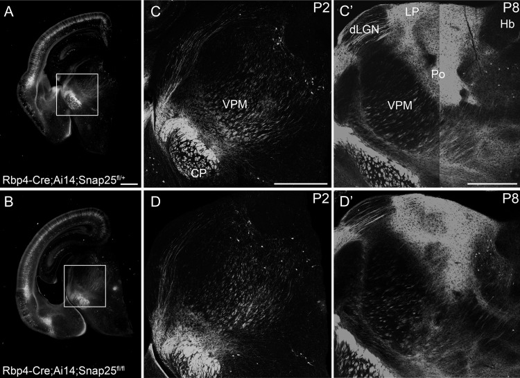
In vivo Cre-expression allows normal development of long-range axonal projections in control and “silenced” Rbp4-Cre expressing cortical L5 projection neurons. Epifluorescent (A, B) and laser scanning confocal microscope tiled images of Rbp4-Cre;Ai14;Snap25fll+ or Rbp4-Cre;Ai14;Snap25fl/fl brains. (A, B) images of P2 brains from Rbp4-Cre;Ai14;Snap25fll+ or Rbp4-Cre;Ai14;Snap25fl/fl mice, demonstrating presence of tdTom+ cells in the cortex, and tdTom+ fibers projecting subcortically. Boxed region enlarged in (C, D). (C, D) tiled confocal images of tdTom+ fibers in the thalamus and cerebral peduncle (CP) at P2. While projections in the cerebral peduncle are already very abundant and dense, projections to higher order thalamic nuclei such as posterior (Po) nucleus are barely detectable at this age. (C′, D′) tiled confocal images of tdTom+ fibers in the thalamus and cerebral peduncle at P8. By this time point, tdTom+ axons course through dorsal lateral geniculate nucleus (dLGN) and form dense terminal arbourisations in Po and lateral posterior nucleus (LP). The subcerebral projections extend within the cerebral peduncle. The pattern, intensity and fasciculation patterns appear identical in the Rbp4-Cre;Ai14;Snap25fll+ and Rbp4-Cre;Ai14;Snap25fl/fl brains. Structures normally devoid of L5 fibers such as habenula do not receive inappropriate innervation in the Rbp4-Cre;Ai14;Snap25fl/fl brain. Note the absence of tdTomato-expression in the dentate gyrus at P2. Scale bars = 500 μm. Abbreviations: CP, cerebral peduncle; dLGN, dorsal lateral geniculate nucleus; Hb, habenula; LP, lateral posterior nucleus; Po, posterior nucleus; VPM, ventral posterior medial nucleus.

