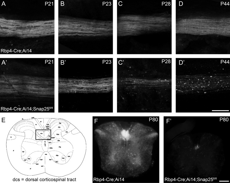
Figure 3.
Rbp4Cre-mediated excision of Snap25fl results in axon degeneration in the spinal cord. Laser scanning confocal microscope (A–D) or epifluorescent (F) images of Rbp4-Cre;Ai14;Snap25fl/+ or Rbp4-Cre;Ai14;Snap25fl/fl brains. (A,A′) at P21, the tdTomato+ axons in longitudinal flat-mounts of the dorsal column of spinal cord (cervical level) from Rbp4-Cre-expressing corticospinal neurons are indistinguishable in Rbp4-Cre;Ai14;Snap25fl/+ and Rbp4-Cre;Ai14;Snap25fl/fl mice. (B,B′) By P23, the tdTomato+ axons in the dorsal column show the first signs of large swellings in Rbp4-Cre;Ai14;Snap25fl/fl mice, but not in controls (C,C′) by the end of the fourth postnatal week, tdTomato+ axons in the dorsal column are reduced in density and the remaining axons show clear signs of discontinuities and axonal swellings in Rbp4-Cre;Ai14;Snap25fl/fl mice, but axons in control are indistinguishable from those at P21. (D,D′) by P44 no continuous axons are present in the dorsal column of the Rbp4-Cre;Ai14;Snap25fl/fl mouse, but residual tdTomato+ punctae remain. Control axons remain normal. (E) schematic diagram (Allen Brain Atlas) of the cross-section of the spinal cord at the cervical level, with boxed area indicating the region from which images in panels (F and F′) are taken. (F,F′) at P80 it is evident in cross-sections of the spinal cord, that there are no tdTomato+ axons remaining in the dorsal column of Rbp4-Cre;Ai14;Snap25fl/fl mice. Scale bars = 100 μm (A–D) and 200 μm (F).

