Fig. 2.
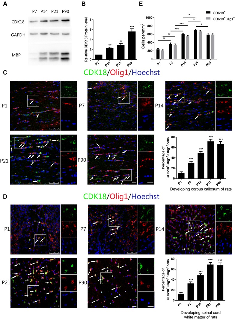
CDK18 is dynamically expressed during development. A, B Western blots (A) and analysis (B) of CDK18 expression in the corpus callosum from P7, P14, P21, and P90 rats. C, D Immunohistochemistry and quantitative analysis of CDK18 expression (green) in Olig1-positive cells (red) in the corpus callosum (C) and white matter of spinal cord (D) from P1, P7, P14, P21, and P90 rats (arrows, cells with co-localization; scale bars, 20 μm). E Quantitative analysis of the density of CDK18 + Olig1 + double-positive and CDK18 + cells throughout postnatal development in spinal cord white matter. Results are expressed as mean ± SEM; *P < 0.05, **P < 0.01, ***P < 0.001, one-way ANOVA with Tukey’s post hoc test; n = 4/group.

