Fig. 5.
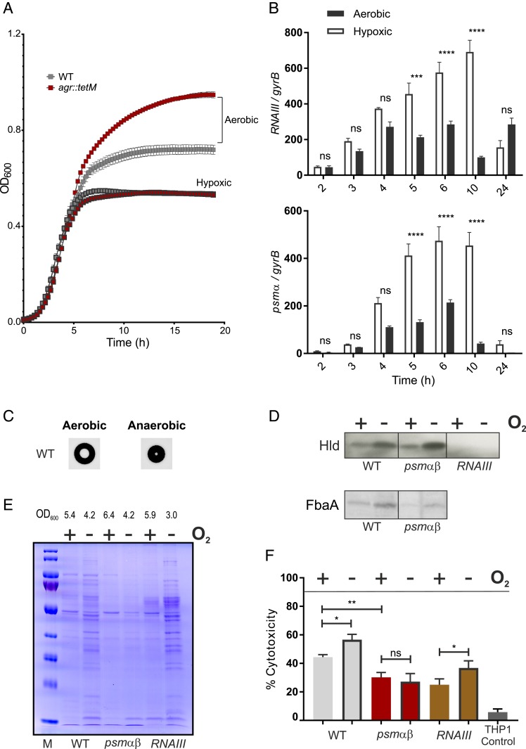
Increased Agr activity in low oxygen environments. (A) Growth of HG001 and agr::tetM under aerobic and hypoxic environments. Error bars represent SD (n = 3). (B) Bacteria were harvested at different time points (2–6, 10, 24 h) during growth. Total RNA from strain HG001 was isolated. RNAIII or psmα mRNA was quantified by qRT-PCR with reference to gyrB. Error bars indicate SEM (n = 3). Statistical significance determined by repeated measures two-way ANOVA with Bonferroni’s posttest. (C) Colony morphology and hemolysis zone of HG001 grown on blood agar under aerobic and anaerobic environments. (D–F) Bacterial supernatants from cultures (HG001, psmα psmβ, and RNAIII) grown either aerobically or under hypoxic conditions were analyzed by (D) Western blot for Hld and aldolase (FbaA) and (E) SDS-PAGE for extracellular proteome (loading control for Western blots). The final bacterial yield in each culture (OD600) is indicated. (F) Bacterial supernatants were analyzed for a cytotoxic potential against THP1 macrophages. Percentage of cytotoxicity shown was normalized to the Triton control. Error bars indicate SD (n = 6). Statistical significance determined by one-way ANOVA with Tukey’s posttest. *P ≤ 0.05, **P ≤ 0.01, ***P ≤ 0.001, and ****P ≤ 0.0001.

