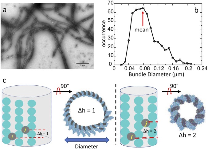
Fig. 4.
Limited diameter of actin bundles. (A) Electron micrograph of negative-stained CaMKIIβ/actin bundles. (B) Diameter distribution of actin bundles in the experiment. (C) Actin forms a curved surface in simulations due to the helical twist of actin filament. The curvature depends on the difference in the number of registers where adjacent CaMKII holoenzymes bind, called “∆h.” If all CaMKII holoenzymes pack with the same pattern, the curved surface will form a barrel. The outer boundary of the barrel dictates the diameter of actin bundles. CaMKII particles are represented by gray circles. Actin subunits are represented by cyan circles.

