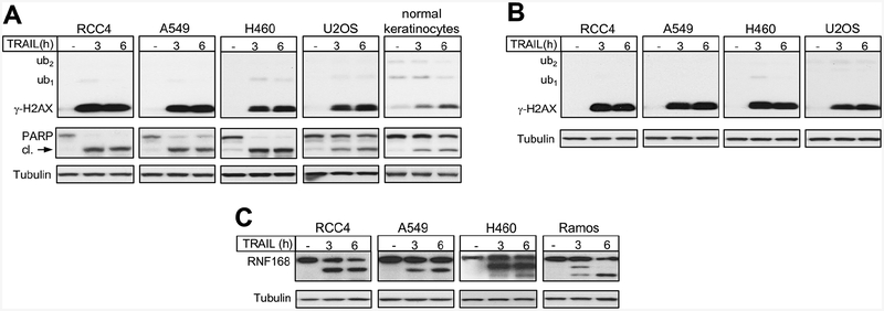
Figure 6. γ-H2AX formation in TRAIL-induced apoptosis.
(A) γ-H2AX formation and PARP cleavage in cells treated with 0.1 μg/ml TRAIL for 3 or 6 h (cl. – cleaved). Semi-dry transfer (buffer with 12% ethanol) from 12% gel and polyclonal antibodies Ab #1 for γ-H2AX were used. (B) Samples from panel A were run on 12% gel, proteins were wet transferred overnight (pH 8.8 buffer with 12% ethanol) and membranes were probed with γ-H2AX polyclonal antibodies Ab #3. (C) Cleavage of RNF168 in cells undergoing TRAIL-induced apoptosis (0.1 μg/ml TRAIL for 3 or 6 h).

