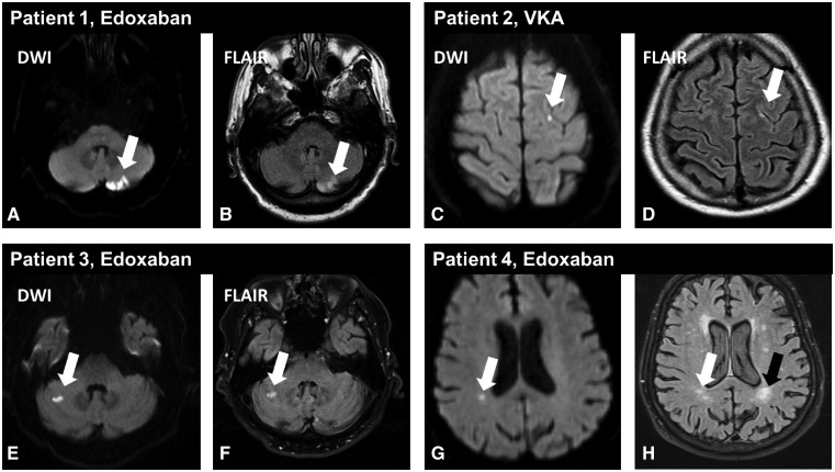
Figure 2.
Examples of acute cerebral microembolism after atrial fibrillation ablation in the magnetic resonance imaging sub-study. Acute cerebral microembolism are depicted by diffusion-weighted imaging (white arrows in A, C, E, G) with corresponding demarcation on fluid-attenuated inversion recovery images (white arrows in B, D, F, H) suggesting an appearance over 24 h ago. (A and B) A larger clinically asymptomatic left cerebellar embolism in an asymptomatic patient randomized to edoxaban. The fluid-attenuated inversion recovery images also revealed chronic white matter hyperintensities (black arrow in H). DWI, diffusion-weighted imaging; FLAIR, fluid-attenuated inversion recovery.

