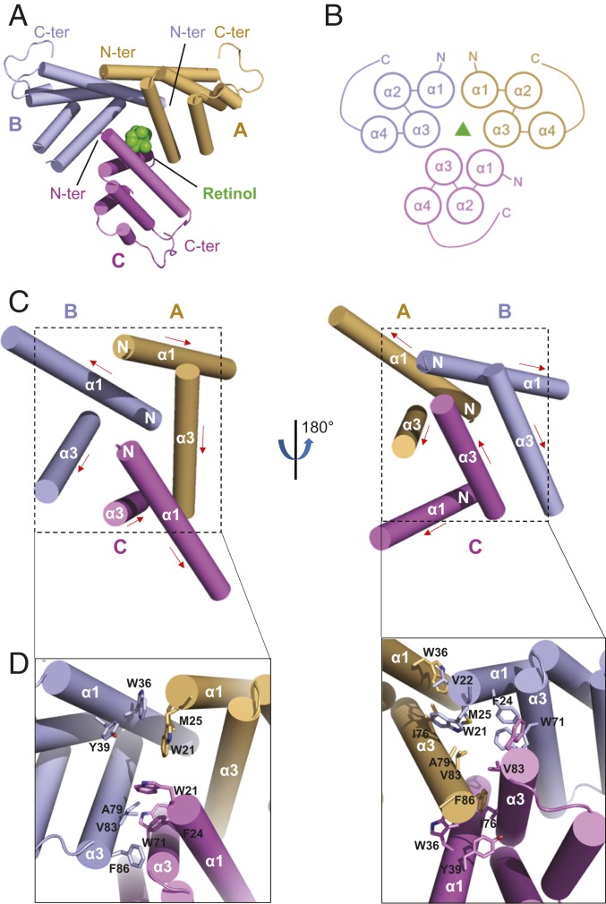
Fig. 3.
Structural details of the SAA3 trimer assembly. (A) Top view of the assembly of SAA3 trimers. Three SAA3 chains are labeled and shown in different colors as indicated. The α-helices are depicted as cylinders, and the bound retinol is shown in green. (B) Topology diagrams of the SAA3 trimer assembly. (C) Helices involved in the assembly of SAA3 trimer shown in 2 different orientations. Three SAA3 molecules are shown in different colors, and helices α1 and α3 from each molecule are labeled. The red arrow indicates the direction from the N terminus to the C terminus. “N” represents the N terminus of each protomer. (D) Amino acids involved in assembly of the SAA3 trimer. Side chains are shown as sticks.

