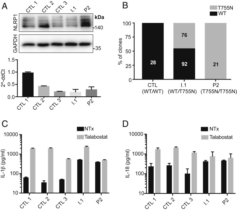
Fig. 3.
Patient-derived keratinocytes show normal expression of NLRP1 protein, baseline inflammasome activation, and unresponsiveness to further NLRP1 activation. (A) Western blot (Top) and qPCR (Bottom) of NLRP1 expression in keratinocytes from P2, heterozygous father (I.1), and 3 controls. The image is representative of 3 independent experiments. (B) Relative expression of NLRP1 WT, T755N transcripts as assessed by TA cloning and Sanger sequencing of an NLPR1 cDNA from keratinocytes from control (CTL), heterozygous father (I.1), and P2. (Inset) Numbers correspond to the number of unique clones sequenced. (C) IL-1β ELISA of supernatants from keratinocytes that were untreated or treated with 3 μM of talabostat (Val-boroPro) for 16 h. (D) IL-18 ELISA of supernatants from keratinocytes that were untreated or treated with 3 μM of talabostat for 16 h. Bars represent mean ± 1 SD. The results are representative of 3 independent experiments.

