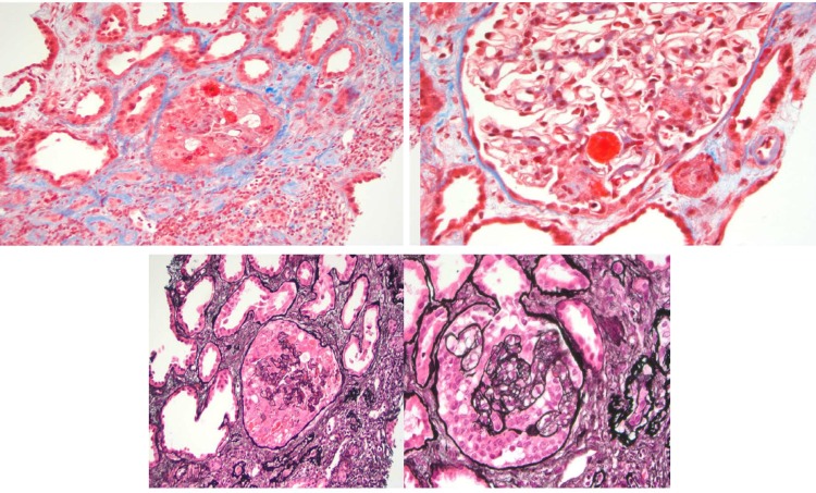
Figure 1.
Light microscopy of glomeruli with thrombotic microangiopathic process and mesangial cell proliferation. Tuft necrosis, rare crescents and mesangiolysis. Forty per cent tubular atrophy and interstitial fibrosis. Immunofluorescence: C3 1+ (focal and granular) and IgM 1+ (focal and granular).

