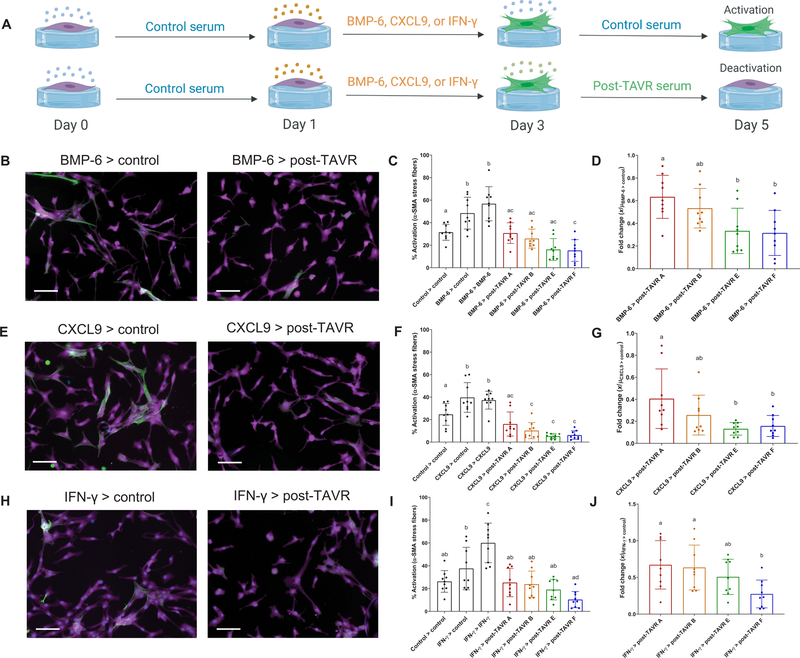
Fig. 3. Pre-TAVR serum factors (BMP-6, CXCL9, and IFN-γ) mediate myofibroblast activation.
(A) Schematic of valvular activation experiments with BMP-6, CXCL9, and IFN-γ (created with BioRender). Representative images of porcine VICs treated initially with (B) BMP-6, (E) CXCL9, or (H) IFN-γ for 2 days (left column) and then treated with either control media or post-TAVR serum (right column). Stains: green, α-SMA; magenta, cytoplasm; blue, nuclei. Scale bars, 100 µm. Percentage of activated VICs initially treated with (C) BMP-6, (F) CXCL9, or (I) IFN-γ and deactivated with post-TAVR serum from four patients. Patient-specific fold changes in post-TAVR deactivation in VICs initially activated with (D) BMP-6, (G) CXCL9, or (J) IFN-γ. Groups with different letters indicate statistical significance (n = 9 measurements per group, means ± SD shown, one-way ANOVA with Tukey posttests, P < 0.05).

