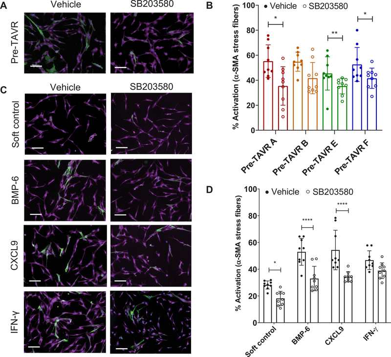
Fig. 4. Validation of p38 MAPK signaling in mediating myofibroblast activation on soft hydrogels in the presence of various factors.
(A) Representative images of porcine VICs treated with pre-TAVR serum in the absence or presence of 20 µM SB203580. Stains: green, α-SMA; magenta, cytoplasm; blue, nuclei. Scale bars, 100 µm. (B) VIC activation in pre-TAVR serum (n = 4 patients) in the absence or presence of SB203580 (n = 9 measurements per group, means ± SD shown, unpaired t test for each patient, *P < 0.05, **P < 0.01). (C) Representative images of VICs treated with BMP-6, CXCL9, and IFN-γ in the absence or presence of 20 µM SB203580. Stains: green, α-SMA; magenta, cytoplasm; blue, nuclei. Scale bars, 100 µm. (D) VIC activation in BMP-6, CXCL9, and IFN-γ in the absence or presence of SB203580 (n = 9 measurements per group, means ± SD shown, two-way ANOVA with Tukey posttests, *P < 0.05, ****P < 0.001).

