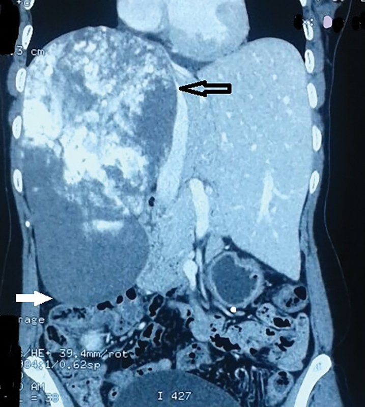
Fig. 1.

Contrast enhanced CT scan demonstrates the huge size of hemangioma which occupies almost the entire right lobe. CT, computed tomography. (Open black arrow shows the medial extend and the solid white arrow shows the inferior extent of the hemangioma).
