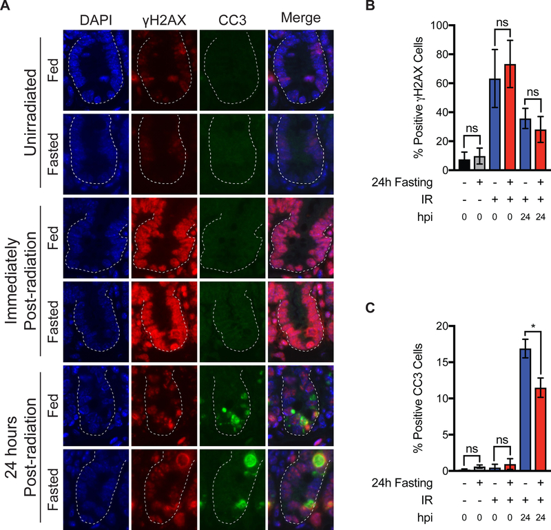
Figure 3:
Effects of pre-radiation fasting on IR-induced DNA damage and apoptosis. (A) C57Bl/6J mice were allowed to feed ad libitum or were fasted for 24 h. Unirradiated SI tissues were harvested at this time. Other cohorts were radiated with TA-XRT (11.5Gy) and SI tissues harvested either immediately or 24 h after radiation (hpi= hours post irradiation). SI tissues (jejunum) were analyzed for γH2AX and cleaved caspase-3 (CC3) by immunofluorescence staining. Representative images are shown. Scale bars, 10 μm. Magnification, 20x. (B) Positive γH2AX cells per crypt were quantified (30 crypts per mouse, n=4 mice per treatment group and mean per treatment plotted. ns= not significant by student’s t-test. Error bars are ± SEM. (C) Positive cleaved caspase-3 cells per crypt were quantified (30 crypts per mouse, n=4 mice per treatment group and mean per treatment plotted. ns= not significant; *P<0.05 by student’s t-test. Error bars are ± SEM.

